
3D/4D ultrasound baby (pregnancy)
During your pregnancy checks, we can use various methods to assess whether everything is in order or whether there are any risks. Ultrasound is the only method that allows us to directly visualize the unborn child in the uterus.
SAFETY
Ultrasound is safe; it has been used in pregnancy for almost 40 years, and no direct harmful effect on the child or mother has ever been demonstrated. With the low sound energies commonly used today, no harm is expected to the expectant child.
THE BENEFITS OF ULTRASOUND
Ultrasound can be used to answer important questions that are important for the further management of the pregnancy:
In the first trimester (10-13 weeks), we can determine the integrity and age of the pregnancy. This is of great importance, for example, to be able to detect reduced growth in late pregnancy. Multiple births can also be detected with a high degree of certainty, and we can also detect a number of severe malformations at this early age. By measuring the nuchal skin, we gain information about a possible chromosomal disorder such as Down syndrome (mongolism).
In the second trimester (20-23 weeks), we assess the baby's growth and amniotic fluid volume, both important indicators of normal development. Important malformations of the head, brain, spine, heart, kidneys, stomach and limbs can be detected. Likewise, the fit of the placenta is assessed because a deep fit or complete covering of the cervix can lead to dangerous bleeding later in pregnancy.
In the third trimester (30-34 weeks), the focus is on fetal growth. A normal-sized baby with normal amniotic fluid levels indicate a functioning placenta. The baby should now have turned in the cranial position to allow for a normal delivery. In addition, some malformations that are important for optimal care of the child after birth can only be detected at this late stage.
VALUE OF ULTRASOUND
If the ultrasound findings are normal, there is a high probability that everything is fine with your child. However, it is not possible to guarantee a perfectly healthy child. Ultrasound is very good (90% accuracy) at detecting very severe fetal problems that may not allow the child to survive. Ultrasound is fairly good (75% accuracy) at detecting problems that require intensive care and therapy. However, ultrasound is more moderately useful (30% accuracy) for detecting minor malformations such as supernumerary fingers or insignificant heart defects.
A normal ultrasound finding has an impact on the further care of the pregnancy and can also greatly reassure you. If a problem is detected, the ultrasound can provide us with important information on which to base decisions; you may be able to prepare for the birth of a sick child, the birth can be planned at a center suitable for that purpose, and occasionally a sick child can be effectively treated during pregnancy.
Routine ultrasounds at 10-13 and 20-23 weeks, as well as other ultrasounds on indication (i.e., when there are risks or suspicions), are covered by health insurance.
The ultrasound is a prenatal examination in the sense of the Law on Human Genetic Testing. Depending on the results of the ultrasound, follow-up examinations such as amniocentesis may be indicated. An unexpected result or evidence of a serious fetal disorder could place you in an ethical decision-making conflict ("should I continue the pregnancy or terminate it?"). In prenatal examinations, there is a right not to know, which is why you should let us know if you do not want an ultrasound for personal reasons. If you are unclear or have any questions, we will be happy to give you more information, and we can arrange for independent expert advice.
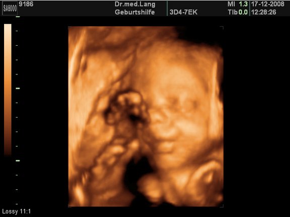
3D/4D ULTRASOUND
Thanks to the latest technology, it is possible to display ultrasound examinations of the child in 3D-4D format. The 3D format shows the child's head in spatial representation (the usual 2D format shows only the profile of the head). The optimal time for the 3D-4D examination is between the 20th and 32nd week of pregnancy (SSW).
Since this service is not a diagnostic examination (diagnosis of malformation), it is not covered by health insurance as a mandatory service. If you wish to have the 3D-4D examination, we will charge you CHF 250 for this additional service. This additional service includes the following package, which is not reimbursed by the health insurance:
- USB stick with Gyné logo (please bring to all consultations);
- 3D/4D images throughout the pregnancy, which are saved on the USB stick. The prerequisite is a favorable position of the unborn child and sufficient amniotic fluid to be able to map the body contours;
- Electronic copy of the medical history in PDF format, updated at each consultation (practical when traveling).
Contact
GYNÉ LANG
Kohlrainstrasse 10
8700 Küsnacht (Zurich)
Phone +41 44 912 25 25
praxis@gynelang.ch
Opening hours
| Monday | 08.00 - 12.00 | 13.30 - 16.30 |
| Tuesday | 08.00 - 12.00 | 13.30 - 16.30 |
| Wednesday | 08.00 - 15.00 |
| Thursday | 08.00 - 12.00 | 13.30 - 16.30 |
| Friday | 08.00 - 15.00 |